EPJ E Highlight - Shocking embryonic limbs into shape
- Details
- Published on 18 September 2019
Electrical stimulation of early chicken embryos has shed light on the process through which the limbs of all vertebrates are formed.
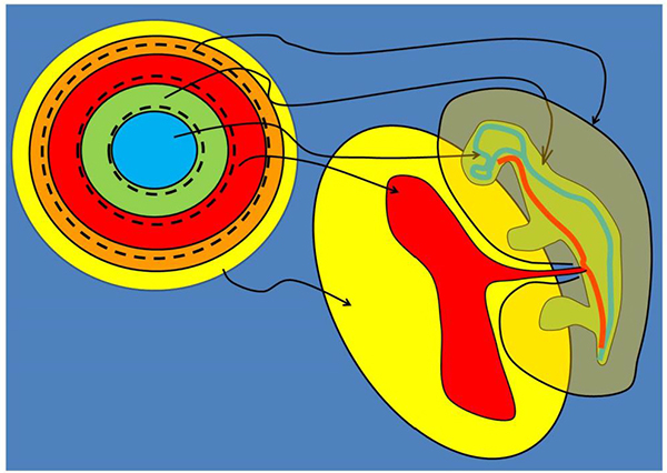
Every vertebrate, whatever its eventual form, starts embryonic life in the same way – as a hollow ball or disc of cells called a blastula. In theory, knowing the mechanism through which the blastula is formed into the shape of an animal could help correct defects and even, one day, regenerate body parts. But evolution and genetics are of little help in understanding this process. Now, however, Vincent Fleury and Ameya Vaishnavi Murukutla from Université Paris Diderot, Paris, France have used experiments with chicken embryos to propose a mechanism for vertebrate limb formation. These findings have been published in the journal EPJ E.Fleury first suggested in 2005 that animal morphogenesis starts by parts of the round blastula being pulled out and deformed in a process that could be thought of as 'string pulling'. His group later showed that the blastula consists of a series of concentric rings, like Russian dolls, and that each forms a different part of the animal.
The novelty of the current study arises from the use of electric shocks to trigger limb formation much more suddenly and rapidly than in natural embryogenesis. The principle behind this is the same phenomenon of bioelectricity that was pioneered by the eighteenth-century Italian physician Galvani, who showed that an electric shock would stimulate even dead muscles into motion. In this study, the researchers extracted early chicken embryos from eggs, incubated them, applied an electric shock and observed them through a powerful microscope. The shock greatly accelerated the formation of the limb and tail buds. Therefore, Fleury and Murukutla were able to see the concentric rings making up the blastula deform in a cascade, with each one's deformation inducing the next to change shape. The authors suggest that knowledge of this phenomenon might eventually lead to the electrical engineering of the tissues of living animals, or of human patients.
V. Fleury and A. V. Murukutla (2019), Electrical stimulation of developmental forces reveals the mechanism of limb formation in vertebrate embryos, Eur. Phys. J. E 42:104. DOI 10.1140/epje/i2019-11869-8