EPJ E Highlight - How do vertebrates take on their form?
- Details
- Published on 16 February 2015
A simple physical mechanism that can be assimilated to folding, or buckling, means that an unformed mass of cells can change in a single step into an embryo organized as a typical vertebrate. This is the main conclusion of work by a team involving physicists from the Laboratoire Matière et Systèmes Complexes (CNRS/Université Paris Diderot) and a biologist from the Laboratoire de Biologie du Développement (CNRS/UPMC).
Thanks to microscopic observations and micromechanical experiments, the scientists have discovered that the pattern that guides this folding is present from the early stages of development. The folds that will give a final shape to the animal form along the boundaries between cell territories with different properties. This work has shed light on the mechanism for the formation of vertebrates and thus how they appeared during evolution. These findings have just been published in EPJ E.
How has evolution produced a structure as complicated as a vertebrate, organized along an anterior-posterior axis, marked dorsally by the nervous system and ventrally by the digestive tract, and displaying almost perfect left-right symmetry? And how, during embryonic development, does this develop from a mass of round cells into an organized embryo? By working on chicken embryos, a team involving physicists and a biologist has managed to explain this transition by means of a relatively simple physical mechanism.
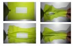
The scientists worked on chicken embryos because at this development stage, they constitute the model that is closest to human embryos. Furthermore, its flat, disk-shaped structure facilitates the observation and modeling of cell movements. A chicken embryo is made up of four concentric rings. Under the microscope, each ring looks like a series of cells of homogenous size; their size increases from the center towards the peripheral rings, with a "stepped" change from one ring to another1. Not only will these cellular domains form different tissues (nervous, muscle, digestive, etc.) but, as discovered by the scientists when filming development of the embryo, it always folds at the boundary between two neighboring rings, as from the second day of its development. These folds will result in a three-dimensional shape, typical of vertebrates.
By measuring the stiffness of the tissues, the scientists were then able to confirm that these boundaries between cell domains display an elastic contrast. The stiffness becomes increasingly marked when the cells are smaller, towards the center of the embryo. Thus as soon as adequate force is applied, the softer, peripheral regions (flanks) "naturally" wrap themselves around the central, stiffer region (the future central nervous system). The force in question is generated by the migration of certain cells, which stretches the embryo lengthwise.
These findings thus offer an explanation for the coupling of cell differentiation and morphogenesis (acquisition by the embryo of its shape), so that a well-formed animal containing territories with different and physically separated functions, emerges "naturally". Understanding this process fills a conceptual gap between a shapeless mass of cells and an "animal archetype", and sheds new light on how vertebrates have emerged during evolution.
Buckling along boundaries of elastic contrast as a mechanism for early vertebrate morphogenesis. Vincent Fleury, Nicolas R. Chevalier, Fabien Furfaro et Jean-Loup Duband (2015), Eur. Phys. J. E 38: 6, DOI 10.1140/epje/i2015-15006-7