EPJ D Highlight - Fragmenting ions and radiation sensitizers
- Details
- Published on 03 September 2019
A new study using mass spectrometry is helping piece together what happens when DNA that has been sensitized by the oncology drug 5-fluorouracil is subjected to the ionising radiation used in radiotherapy.
The anti-cancer drug 5-fluorouracil (5FU) acts as a radiosensitizer: it is rapidly taken up into the DNA of cancer cells, making the cells more sensitive to radiotherapy. However, little is known about the precise mechanism through which radiation damages cells. A team of scientists led by Peter van der Burgt at the National University of Ireland in Maynooth, Ireland have now used mass spectrometry to shed some light on this process; their work was recently published in EPJ D. A full understanding of this process could ultimately lead to new ways of protecting normal tissues from the radiation damage caused by essential cancer treatments.
Radiotherapy involves delivering radiation, in the form of X-rays, to the cancer site. The X-rays or other high-energy particles collide with the molecules they encounter, knocking off electrons to form positively-charged ions, and these ions and electrons in turn react with other molecules in the cells: this can kill cancer cells, but can also cause short- and long-term damage to others.
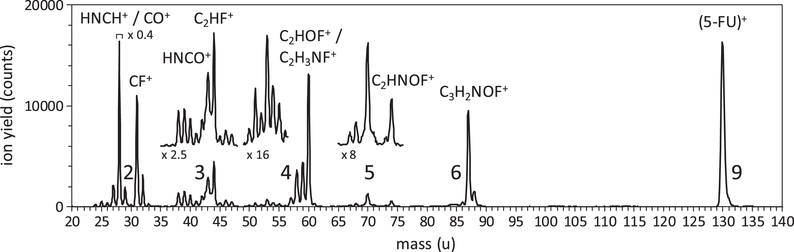
5-fluorouracil resembles the bases in normal DNA but is more sensitive to radiation, both on its own and when incorporated into DNA. Van der Burgt and his colleagues investigated this sensitivity by producing a stream of 5FU particles in a mass spectrometer and colliding them with a beam of radiation. The spectrometer was then used to determine which ions were formed in the collisions by measuring their mass-to-charge ratio. Two sets of experiments were carried out: one using electrons as the radiation source, and the other using high-intensity photons.
Increasing the energy of the radiation increased the number and type of ions that were formed; the researchers identified many different fragment ions and determined the radiation level at which each was first formed. These included some ions that were absent from the spectrum of the natural base, uracil, and some that had not been observed in this type of experiment before. Undoubtedly, similar processes take place in the tissues of patients undergoing radiotherapy.
P.J.M. van der Burgt, M.A. Brown, J. Bockova, A. Rebelo, M. Ryszka, J-C. Poully and S. Eden (2019) Fragmentation processes of ionized 5-fluorouracil in the gas phase and within clusters, European Physical Journal D 73: 184, DOI: 10.1140/epjd/e2019-100107-7